Quantifying CA1 Dorsal Hippocampal Pyramidal Cells in rats : rules to light microscope based estimation
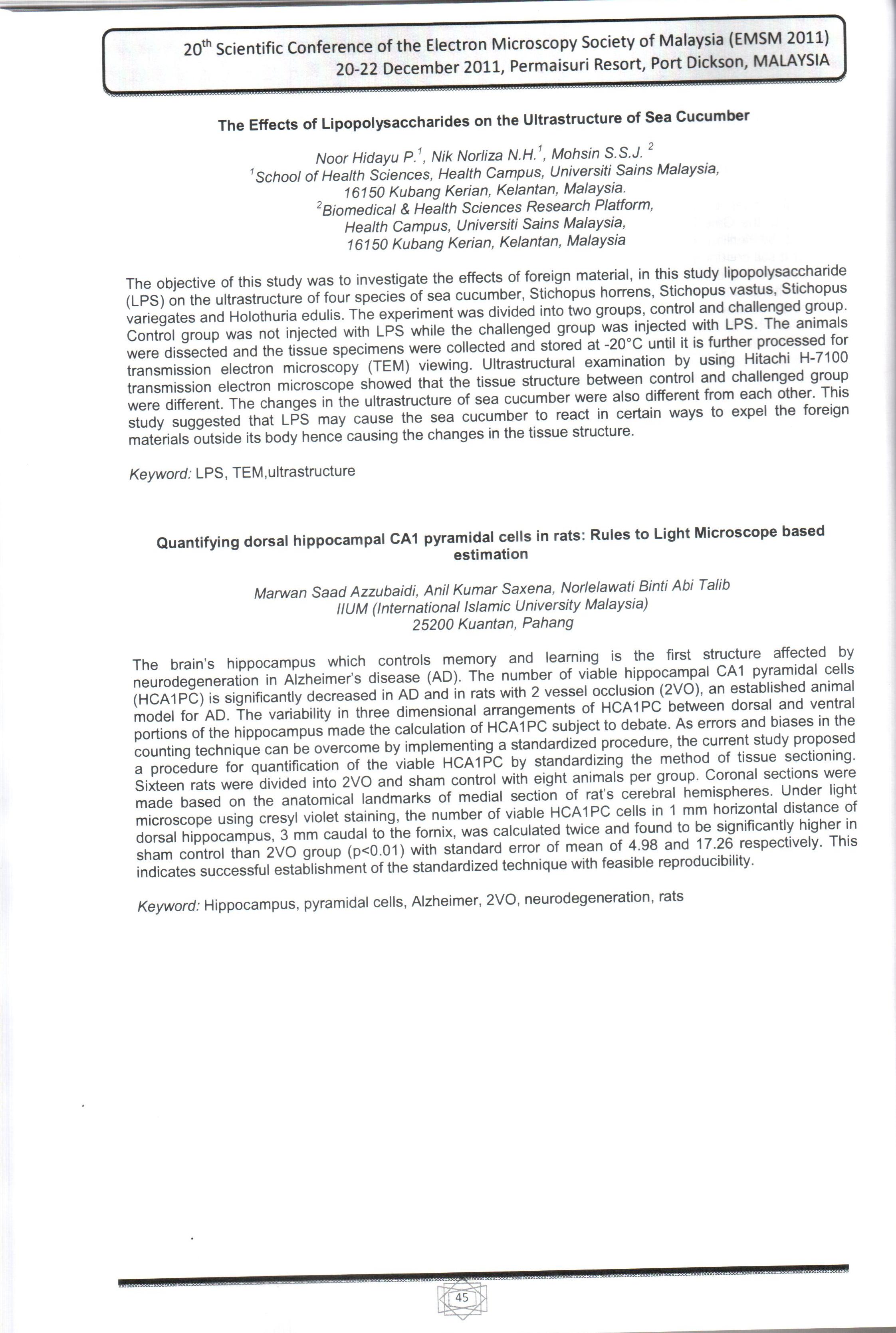
The brain’s hippocampus which controls memory and learning is the first structure affected by neurodegeneration in Alzheimer’s disease (AD). The number of viable hippocampal CA1 pyramidal cells (HCA1PC) is significantly decreased in AD and in rats with 2 vessel occlusion (2VO), an established anima...
| Main Authors: | , , |
|---|---|
| 格式: | Proceeding Paper |
| 語言: | English English English |
| 出版: |
2011
|
| 主題: | |
| 在線閱讀: | http://irep.iium.edu.my/13125/1/EMSM_cover_page.jpg http://irep.iium.edu.my/13125/2/EMSM_abstract_book_pp45.jpg http://irep.iium.edu.my/13125/3/Hippocampal_CA1_pyramidal_cell_calculation_standerdization.pdf |
| _version_ | 1825645376654278656 |
|---|---|
| author | Azzubaidi, Marwan Saad Saxena, Anil Kumar A.Talib, Norlelawati |
| author_facet | Azzubaidi, Marwan Saad Saxena, Anil Kumar A.Talib, Norlelawati |
| author_sort | Azzubaidi, Marwan Saad |
| collection | IIUM |
| description | The brain’s hippocampus which controls memory and learning is the first structure affected by neurodegeneration in Alzheimer’s disease (AD). The number of viable hippocampal CA1 pyramidal cells (HCA1PC) is significantly decreased in AD and in rats with 2 vessel occlusion (2VO), an established animal model for AD. The variability in three dimensional arrangements of HCA1PC between dorsal and ventral portions of the hippocampus made the calculation of HCA1PC subject to debate. As errors and biases in the counting technique can be overcome by implementing a standardized procedure, the current study proposed a procedure for quantification of the viable HCA1PC by standardizing the method of tissue sectioning. Sixteen rats were divided into 2VO and sham control with eight animals per group. Coronal sections were made based on the anatomical landmarks of medial section of rat’s cerebral hemispheres. Under light microscope with cresyl violet staining, the number of viable HCA1PC cells in 1 mm horizontal distance of dorsal hippocampus, 3 mm caudal to the fornix, was calculated twice and found to be significantly higher in sham control than 2VO group (p<0.01) with standard error of mean of 4.98 and 17.26 respectively. This indicates successful establishment of the standardized technique with feasible reproducibility.
Keywords: hippocampus, CA1 area, pyramidal cells, rat
|
| first_indexed | 2024-03-05T22:46:48Z |
| format | Proceeding Paper |
| id | oai:generic.eprints.org:13125 |
| institution | International Islamic University Malaysia |
| language | English English English |
| last_indexed | 2024-03-05T22:46:48Z |
| publishDate | 2011 |
| record_format | dspace |
| spelling | oai:generic.eprints.org:131252012-12-17T06:18:22Z http://irep.iium.edu.my/13125/ Quantifying CA1 Dorsal Hippocampal Pyramidal Cells in rats : rules to light microscope based estimation Azzubaidi, Marwan Saad Saxena, Anil Kumar A.Talib, Norlelawati R Medicine (General) The brain’s hippocampus which controls memory and learning is the first structure affected by neurodegeneration in Alzheimer’s disease (AD). The number of viable hippocampal CA1 pyramidal cells (HCA1PC) is significantly decreased in AD and in rats with 2 vessel occlusion (2VO), an established animal model for AD. The variability in three dimensional arrangements of HCA1PC between dorsal and ventral portions of the hippocampus made the calculation of HCA1PC subject to debate. As errors and biases in the counting technique can be overcome by implementing a standardized procedure, the current study proposed a procedure for quantification of the viable HCA1PC by standardizing the method of tissue sectioning. Sixteen rats were divided into 2VO and sham control with eight animals per group. Coronal sections were made based on the anatomical landmarks of medial section of rat’s cerebral hemispheres. Under light microscope with cresyl violet staining, the number of viable HCA1PC cells in 1 mm horizontal distance of dorsal hippocampus, 3 mm caudal to the fornix, was calculated twice and found to be significantly higher in sham control than 2VO group (p<0.01) with standard error of mean of 4.98 and 17.26 respectively. This indicates successful establishment of the standardized technique with feasible reproducibility. Keywords: hippocampus, CA1 area, pyramidal cells, rat 2011-12-20 Proceeding Paper NonPeerReviewed application/pdf en http://irep.iium.edu.my/13125/1/EMSM_cover_page.jpg application/pdf en http://irep.iium.edu.my/13125/2/EMSM_abstract_book_pp45.jpg application/pdf en http://irep.iium.edu.my/13125/3/Hippocampal_CA1_pyramidal_cell_calculation_standerdization.pdf Azzubaidi, Marwan Saad and Saxena, Anil Kumar and A.Talib, Norlelawati (2011) Quantifying CA1 Dorsal Hippocampal Pyramidal Cells in rats : rules to light microscope based estimation. In: 20th Scientific Conference of the Microscopy society of Malaysia, 20th - 22nd December 2011, Port Disckson, NS.. |
| spellingShingle | R Medicine (General) Azzubaidi, Marwan Saad Saxena, Anil Kumar A.Talib, Norlelawati Quantifying CA1 Dorsal Hippocampal Pyramidal Cells in rats : rules to light microscope based estimation |
| title | Quantifying CA1 Dorsal Hippocampal Pyramidal Cells in rats : rules to light microscope based estimation |
| title_full | Quantifying CA1 Dorsal Hippocampal Pyramidal Cells in rats : rules to light microscope based estimation |
| title_fullStr | Quantifying CA1 Dorsal Hippocampal Pyramidal Cells in rats : rules to light microscope based estimation |
| title_full_unstemmed | Quantifying CA1 Dorsal Hippocampal Pyramidal Cells in rats : rules to light microscope based estimation |
| title_short | Quantifying CA1 Dorsal Hippocampal Pyramidal Cells in rats : rules to light microscope based estimation |
| title_sort | quantifying ca1 dorsal hippocampal pyramidal cells in rats rules to light microscope based estimation |
| topic | R Medicine (General) |
| url | http://irep.iium.edu.my/13125/1/EMSM_cover_page.jpg http://irep.iium.edu.my/13125/2/EMSM_abstract_book_pp45.jpg http://irep.iium.edu.my/13125/3/Hippocampal_CA1_pyramidal_cell_calculation_standerdization.pdf |
| work_keys_str_mv | AT azzubaidimarwansaad quantifyingca1dorsalhippocampalpyramidalcellsinratsrulestolightmicroscopebasedestimation AT saxenaanilkumar quantifyingca1dorsalhippocampalpyramidalcellsinratsrulestolightmicroscopebasedestimation AT atalibnorlelawati quantifyingca1dorsalhippocampalpyramidalcellsinratsrulestolightmicroscopebasedestimation |